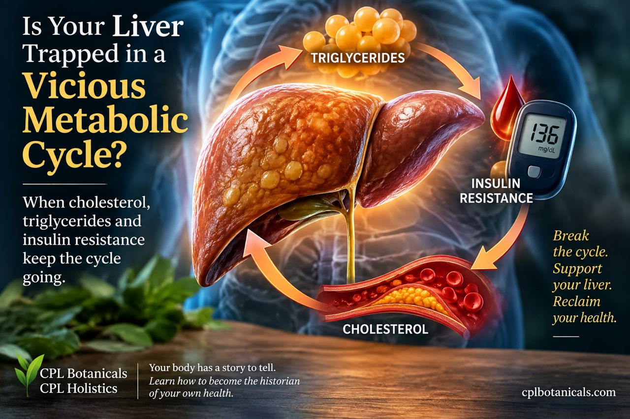
When liver cholesterol and triglycerides show up as concerns on a lab report, food usually gets blamed first. Too much fat, too much sugar, or too many carbohydrates can certainly influence those numbers, but diet is only part of the picture. Your liver is working every minute to decide what to make, what to store, what to burn, and what to send back into the bloodstream. That makes a lipid panel more than a report about what you ate.
Cholesterol, triglycerides, blood sugar, insulin, body fat, and liver fat all intersect through shared metabolic pathways. In insulin resistance, changes can begin in the liver, skeletal muscle, fat tissue, and pancreas long before type 2 diabetes appears on a lab report. NIDDK explains that insulin resistance can occur in muscle, fat, and liver cells and may accompany unhealthy cholesterol and triglyceride levels and fatty liver disease¹. Researchers are now examining another part of that picture: cellular senescence, often called “zombie cells,” and the possibility that these dysfunctional cells help keep metabolic problems going.
Your Liver Is a Metabolic Control Center
Your liver does far more than filter substances or make bile. It helps control glucose, fatty acids, cholesterol, and triglycerides, constantly adjusting those jobs according to the signals it receives. Cholesterol is essential for cell membranes, steroid hormones, and bile acids, so the body cannot simply eliminate it. The liver makes cholesterol, receives it from circulating particles, repackages it, and helps remove excess amounts through bile.
Triglycerides serve mainly as stored energy. Fatty acids can reach the liver from food or body fat, and the liver can also make new fatty acids from excess carbohydrate and other energy sources through de novo lipogenesis. It can burn some fatty acids for energy, store some as triglycerides, or package triglycerides into very-low-density lipoprotein, better known as VLDL, for release into the bloodstream. Research on insulin resistance and lipoprotein metabolism² shows how strongly liver function influences VLDL, triglycerides, LDL, and HDL.
How VLDL Connects Triglycerides to Cholesterol
VLDL particles leave the liver carrying a large amount of triglyceride. As enzymes remove triglycerides from those particles and deliver fatty acids to tissues, the particles become progressively smaller and can eventually contribute to LDL formation. Triglyceride metabolism and cholesterol metabolism therefore function as connected systems rather than two unrelated processes.
When the liver sends out too much VLDL, triglycerides can rise and the entire lipoprotein pattern can change. HDL often falls, while smaller, denser LDL particles can become more common. A review of lipids and lipoproteins in type 2 diabetes³ identifies increased hepatic secretion of triglyceride-rich VLDL and impaired VLDL clearance as central features of diabetic dyslipidemia. A standard lipid panel may therefore show several abnormal values that trace back to overlapping metabolic problems rather than a collection of unrelated dietary mistakes.
Insulin Has a Job in the Liver Too
Most people think of insulin as the hormone that lowers blood sugar, but insulin also helps coordinate liver metabolism. After a meal, rising insulin normally tells the liver to reduce its glucose output and influences how the liver handles fat. Muscle and fat tissue receive insulin’s message as well, helping those tissues take up or store nutrients according to the body’s needs.
With insulin resistance, those signals lose some of their effect. Muscle may take up less glucose, fat tissue may release more fatty acids, and the liver may continue producing glucose when it should be slowing down. At the same time, the insulin-resistant liver can continue making and exporting triglyceride-rich particles. Research on selective hepatic insulin resistance⁴ describes how VLDL overproduction and hypertriglyceridemia develop as insulin signaling becomes disturbed. This helps explain why insulin resistance can affect blood sugar and blood fats at the same time.
Why Triglycerides Often Rise With Insulin Resistance
Insulin normally helps keep stored fat inside adipose tissue after a meal. When fat tissue becomes insulin resistant, more fatty acids can escape into circulation and travel to the liver. Meanwhile, the liver may increase de novo lipogenesis, adding newly made fat to the incoming supply. It must then decide how much of that increasing fuel supply to burn, store, or send back into circulation.
When the liver packages more triglyceride into VLDL, blood triglycerides can climb. Mechanistic research on hepatic VLDL overproduction⁵ links insulin resistance with increased fatty-acid delivery, altered insulin signaling, and greater VLDL output. This helps explain why triglycerides may begin changing while fasting glucose still looks acceptable. A triglyceride value cannot diagnose insulin resistance by itself, but it can add an important clue when it appears alongside rising glucose, increased waist size, fatty liver, lower HDL, or other metabolic changes.
When Fat Starts Accumulating in the Liver
The liver normally contains some fat, but problems arise when more fat arrives or gets made than the liver can burn or export. Triglycerides then accumulate inside liver cells, producing metabolic dysfunction-associated steatotic liver disease, or MASLD. Many people still recognize its older name, nonalcoholic fatty liver disease.
Insulin resistance and MASLD frequently reinforce one another. Insulin-resistant adipose tissue sends more fatty acids toward the liver, while de novo lipogenesis can increase inside the liver itself. As liver fat grows, hepatic insulin signaling can deteriorate further. The 2024 EASL, EASD, and EASO clinical guidelines⁶ emphasize the close relationship among MASLD, type 2 diabetes, obesity, and other cardiometabolic risk factors. Liver fat therefore belongs in the same conversation as triglycerides, glucose, insulin resistance, and cardiovascular risk.
Liver Fat Is More Than Stored Energy
It would be easy to picture fatty liver as a storage problem only, but liver cells experience much more than simple fat accumulation. Chronic nutrient excess can increase oxidative stress, disturb mitochondrial function, alter fatty-acid oxidation, and activate inflammatory pathways. Those changes can affect how liver cells respond to insulin and how effectively they manage incoming and outgoing fuel.
Triglyceride storage itself may sometimes protect cells by locking fatty acids into a less reactive form. The greater concern can come from the broader environment created by continued metabolic overload, harmful lipid intermediates, oxidative stress, and impaired cellular energy handling. As those pressures continue, the liver may struggle with both glucose regulation and lipid management. This is where the cellular-senescence research becomes especially relevant to the liver story.
Where “Zombie Cells” Fit Into This Story
We have already covered the basic biology of senescent cells in earlier articles, so only a short refresher is necessary here. A senescent cell stops dividing but remains metabolically active. Senescence can serve useful purposes, including limiting the growth of damaged cells and participating in tissue repair, so the process itself is not automatically harmful.
Trouble can develop when senescent cells accumulate and persist. Some release inflammatory cytokines, chemokines, enzymes, and other signaling molecules known collectively as the senescence-associated secretory phenotype, or SASP. Those signals can alter neighboring cells and the tissue around them. A review of senescence in type 2 diabetes⁷ describes how senescent cells in metabolic tissues may contribute to pancreatic beta-cell dysfunction, adipose dysfunction, inflammation, and insulin resistance. For this discussion, the important question is what happens when senescence appears in the tissues responsible for managing fuel.
Senescent Liver Cells May Handle Fat Differently
Researchers studying MASLD have found a two-way relationship between lipid accumulation and cellular senescence. Excess fatty acids and metabolic stress can encourage senescence, while persistent senescence can interfere with normal lipid handling. Senescent hepatocytes may increase lipid uptake, develop mitochondrial problems, and eventually lose some capacity for fatty-acid oxidation.
SASP signaling can add another problem by maintaining inflammation inside the liver. A 2026 review focused on senescence and lipid metabolism in MASLD⁸ describes a feedback loop among lipid accumulation, mitochondrial dysfunction, inflammatory signaling, and cellular senescence. The research does not show that senescent liver cells single-handedly cause fatty liver or diabetes. Instead, it suggests that metabolic stress can promote senescence while senescence may make normal lipid handling and insulin responsiveness increasingly difficult.
Fat Tissue Can Spread the Metabolic Problem
Adipose tissue is an active endocrine organ, not an inert storage container. Healthy fat cells store energy after meals and release fatty acids when the body needs them. They also send hormonal and inflammatory signals that affect the liver, muscle, pancreas, and other tissues. When fat cells become dysfunctional, their problems can spread throughout the metabolic system.
Human research has found more senescence in mature adipose cells from people with type 2 diabetes, even after researchers matched participants for age and body mass index. The degree of adipose-cell senescence correlated with whole-body insulin resistance, and the cells showed lower levels of proteins important for insulin action, including GLUT4. The human adipose-cell study published in Diabetes⁹ gives us direct evidence that senescence and insulin resistance can coexist within human metabolic tissue. Dysfunctional fat tissue can then send more fatty acids toward the liver, adding another source of metabolic pressure.
Skeletal Muscle Matters More Than Most People Realize
Skeletal muscle handles a large share of insulin-stimulated glucose disposal after meals. When insulin works properly, muscle cells pull glucose from the bloodstream and either burn it for energy or store it as glycogen. If muscle becomes insulin resistant, the pancreas often compensates by producing more insulin to push the same message harder. That compensation can keep glucose within a normal range for a time, which helps explain why insulin resistance can develop before fasting glucose or A1C reaches the diabetic range.
Human research is also beginning to connect muscle senescence with insulin sensitivity. In a 2025 study using human skeletal-muscle biopsies, researchers found higher senescence markers and impaired insulin-related signaling in participants with obesity. Exercise improved insulin sensitivity while reducing several senescence markers. The human skeletal-muscle study¹⁰ adds another piece to the picture because skeletal muscle plays such a large role in glucose disposal. Insulin resistance increasingly looks like disrupted communication among several metabolic tissues rather than one defective receptor or one malfunctioning organ.
The Pancreas Eventually Feels the Strain
Pancreatic beta cells make insulin, and they often work harder during the early stages of insulin resistance. Higher insulin output can compensate for resistance in muscle, liver, and fat tissue, sometimes for years. Blood glucose may therefore remain below the diabetic range even while the pancreas is producing more insulin to maintain that result.
Chronic metabolic stress can eventually affect the beta cells themselves. Elevated glucose, excessive fatty acids, oxidative stress, and inflammatory signals can all contribute to beta-cell dysfunction, and researchers have identified cellular senescence in pancreatic beta cells as another possible factor. Research on cellular senescence in type 2 diabetes¹¹ describes a possible pathogenic loop in which senescent cells contribute to metabolic dysfunction while diabetes-related changes promote additional senescence. The pancreas may therefore respond to insulin resistance elsewhere while simultaneously facing cellular stress of its own.
Type 2 Diabetes May Promote Senescence Too
The relationship between senescence and diabetes does not run in only one direction. High glucose can accelerate senescence through several cellular pathways, while oxidative stress, altered lipid metabolism, inflammation, and mitochondrial dysfunction can create additional cellular stress. Metabolic disease may therefore create conditions that encourage more senescent cells to accumulate.
At the same time, those cells can release SASP factors that worsen inflammatory signaling and interfere with tissue function. Research on the regulation of cellular senescence in type 2 diabetes¹² discusses hyperglycemia-driven senescence and the possibility that senescence becomes both a consequence and a contributor to diabetes. This two-way relationship gives us a more accurate picture than the oversimplified claim that “zombie cells cause diabetes.” Current evidence points toward a feedback cycle involving metabolic stress, cellular dysfunction, inflammation, and impaired insulin signaling.
What the Newer 2026 Research Adds
A 2026 review pulled together preclinical and human evidence on senescence in type 2 diabetes and reached an important conclusion. Animal studies consistently show that removing certain senescent cells can improve insulin sensitivity, reduce SASP-related inflammation, and preserve beta-cell function in diabetic models. Those findings strengthen the argument that senescent cells can actively participate in metabolic dysfunction rather than simply appearing alongside it.
Human evidence tells a more cautious story. Current trials remain small and short, and researchers have focused heavily on biological markers and safety rather than long-term diabetes outcomes. The 2026 review of cellular senescence and metabolic aging in type 2 diabetes¹³ describes senescence as a biologically plausible contributor to metabolic dysfunction while emphasizing that senolytic treatment remains experimental. We have enough evidence to take the mechanism seriously, but not enough to say that clearing senescent cells will prevent, reverse, or cure type 2 diabetes in people.
What About Senolytics?
Senolytics are compounds designed to selectively eliminate certain senescent cells. In animal models, this approach has produced improvements in insulin sensitivity, inflammation, beta-cell function, and other metabolic measures. Those results have generated understandable interest because researchers may eventually be able to target a process that helps sustain metabolic dysfunction rather than focusing only on its downstream effects.
Human research has not reached that point. Scientists still need to identify which senescent cells cause harm, when removing them helps, how selectively a treatment can target them, and whether benefits last without interfering with useful functions of senescence. A review examining cellular senescence at the intersection of aging and diabetes¹⁴ explains both the therapeutic promise and the unanswered questions surrounding senolytics. For now, supplements marketed as “senolytic” should not be presented as established treatments for insulin resistance or type 2 diabetes.
Why Blood Sugar Alone Does Not Tell the Whole Story
Type 2 diabetes becomes visible through abnormal glucose, but the metabolic disruption often involves far more than glucose. Muscle may take up less glucose, fat tissue may release more fatty acids, the liver may continue producing glucose, and the pancreas may increase insulin output to compensate. Meanwhile, the liver can produce more VLDL, triglycerides can rise, HDL can fall, and liver fat can increase.
That clustering is one reason insulin resistance frequently appears alongside dyslipidemia and fatty liver. Research on diabetic dyslipidemia¹⁵ describes elevated triglycerides, low HDL, altered LDL particles, increased free fatty acids, and low-grade inflammation as interconnected features of insulin-resistant metabolism. Senescence research adds another possible layer by showing how persistent cellular stress and inflammatory signaling may help maintain dysfunction across several organs. Looking only at glucose can therefore miss parts of the metabolic process that have already begun changing.
Your Triglycerides Can Be an Important Clue
A triglyceride result cannot tell you why it is high, and it cannot diagnose insulin resistance on its own. Alcohol intake, genetics, medications, thyroid disease, kidney disease, diet, poorly controlled diabetes, and other factors can also raise triglycerides. Still, triglycerides can provide useful information when clinicians interpret them alongside glucose, A1C, HDL, LDL, waist size, blood pressure, liver findings, medications, and personal history.
In insulin-resistant states, elevated triglycerides frequently accompany increased hepatic VLDL secretion and changes in HDL and LDL particles. That pattern can appear before someone meets the diagnostic criteria for type 2 diabetes. Looking at the pattern does not mean blaming every abnormal lipid value on insulin resistance. It means recognizing that the liver, pancreas, muscle, and adipose tissue participate in the same metabolic conversation, and changes in one part of the system can show up somewhere else.
The Bigger Story Behind the Numbers
Your body does not separate cholesterol, triglycerides, glucose, insulin, and liver fat into different boxes simply because a laboratory report does. The liver receives fatty acids from adipose tissue, responds to insulin from the pancreas, manages glucose, produces and clears lipoproteins, and reacts to inflammatory and cellular stress signals. Muscle affects how much glucose leaves the bloodstream, while adipose tissue affects how much fat reaches the liver.
Cellular senescence does not replace the established explanations for insulin resistance and type 2 diabetes. It adds another possible mechanism within them. The strongest evidence currently shows that senescence can participate in inflammatory signaling, tissue dysfunction, altered lipid handling, and impaired insulin responsiveness, while metabolic stress can encourage even more senescence. That feedback cycle may become an important target for future treatment, but human evidence still needs to catch up with the animal research.
Your body has a story to tell, and laboratory values can become part of that story when we stop viewing them as isolated numbers. Cholesterol, triglycerides, blood sugar, liver fat, and insulin resistance may represent different pages of the same metabolic chapter. Reading those pages together can reveal connections that disappear when attention stays fixed on one result at a time.
Work With Charlotte
When blood sugar, cholesterol, triglycerides, blood pressure, weight changes, energy, and other health concerns get considered one at a time, important patterns can disappear. My naturopathic coaching consultations help you organize your health history, nutrition, lifestyle, symptoms, and laboratory information so you can see connections and prepare better questions for your healthcare providers. The goal is to understand your own patterns and become a more informed participant in your care. Your body has a story to tell. Learn how to become the historian of your own health.
Herbally and Holistically Yours,
Charlotte Lange, CNC
CPL Holistics | CPL BotanicalsReferences
- National Institute of Diabetes and Digestive and Kidney Diseases. Insulin Resistance & Prediabetes.
https://www.niddk.nih.gov/health-information/diabetes/overview/what-is-diabetes/prediabetes-insulin-resistance- Ormazabal V, et al. Pathogenesis of Lipid Disorders in Insulin Resistance: A Brief Review.
https://pubmed.ncbi.nlm.nih.gov/30328521/- Garvey WT, Kwon S, Zheng D, et al. Lipids and Lipoproteins in Patients With Type 2 Diabetes.
https://pubmed.ncbi.nlm.nih.gov/15161808/- Sparks JD, Sparks CE, Adeli K. Selective Hepatic Insulin Resistance, VLDL Overproduction, and Hypertriglyceridemia.
https://pubmed.ncbi.nlm.nih.gov/22796579/- Taghibiglou C, Carpentier A, Van Iderstine SC, et al. Mechanisms of Hepatic Very Low-Density Lipoprotein Overproduction in Insulin Resistance.
https://pubmed.ncbi.nlm.nih.gov/11597827/- European Association for the Study of the Liver, European Association for the Study of Diabetes, European Association for the Study of Obesity. EASL-EASD-EASO Clinical Practice Guidelines on the Management of Metabolic Dysfunction-Associated Steatotic Liver Disease.
https://pubmed.ncbi.nlm.nih.gov/38851997/- Palmer AK, Gustafson B, Kirkland JL, Smith U. Role of Cellular Senescence in Type II Diabetes.
https://pubmed.ncbi.nlm.nih.gov/34363464/- Mastoridou EM, et al. The Interplay Between Cellular Senescence and Lipid Metabolism in the Progression of Metabolic Dysfunction-Associated Steatotic Liver Disease.
https://pmc.ncbi.nlm.nih.gov/articles/PMC12842010/- Gustafson B, Nerstedt A, Spinelli R, Beguinot F, Smith U. Type 2 Diabetes, Independent of Obesity and Age, Is Characterized by Senescent and Dysfunctional Mature Human Adipose Cells.
https://pubmed.ncbi.nlm.nih.gov/36006465/- Podraza-Farhanieh A, Spinelli R, Zatterale F, et al. Physical Training Reduces Cell Senescence and Associated Insulin Resistance in Skeletal Muscle.
https://pubmed.ncbi.nlm.nih.gov/40127780/- Palmer AK, Tchkonia T, LeBrasseur NK, Chini EN, Xu M, Kirkland JL. Cellular Senescence in Type 2 Diabetes: A Therapeutic Opportunity.
https://pubmed.ncbi.nlm.nih.gov/26106186/- Kitada M, Koya D. Regulation of Cellular Senescence in Type 2 Diabetes Mellitus: From Mechanisms to Clinical Applications.
https://pubmed.ncbi.nlm.nih.gov/36872059/- Syed AR, et al. Cellular Senescence and Metabolic Aging in Type 2 Diabetes: Mechanistic Insights and Translational Implications. 2026.
https://pubmed.ncbi.nlm.nih.gov/41948554/- Palmer AK, Gustafson B, Kirkland JL, Smith U. Cellular Senescence: At the Nexus Between Ageing and Diabetes.
https://pubmed.ncbi.nlm.nih.gov/31451866/- Wu L, Parhofer KG. Diabetic Dyslipidemia.
https://pubmed.ncbi.nlm.nih.gov/25242435/
Leave a Reply